Reproduction is a fundamental biological process that ensures the continuation of a species. In mammals like dogs and cats, sexual reproduction involves the fusion of specialized germ cells—spermatozoa from the male and ova from the female—to form a zygote. This process not only perpetuates the species but also introduces genetic diversity, which is essential for adaptation and evolution. The reproductive systems of dogs and cats share many similarities, though there are notable differences that reflect their unique evolutionary paths.
Male Reproductive System
The male reproductive system in dogs and cats is designed to produce, store, and deliver sperm to the female reproductive tract. It consists of several key components, each playing a critical role in the reproductive process.
Testis: The Male Gonad
The testis is the primary male reproductive organ, responsible for two critical functions:
- Spermatogenesis: The production of spermatozoa (sperm) through a process called meiosis, which reduces the chromosome number by half to ensure genetic diversity in offspring.
- Hormone Secretion: The testis secretes testosterone, a hormone that influences spermatogenesis, the development of male secondary sexual characteristics, and male behavioral patterns.
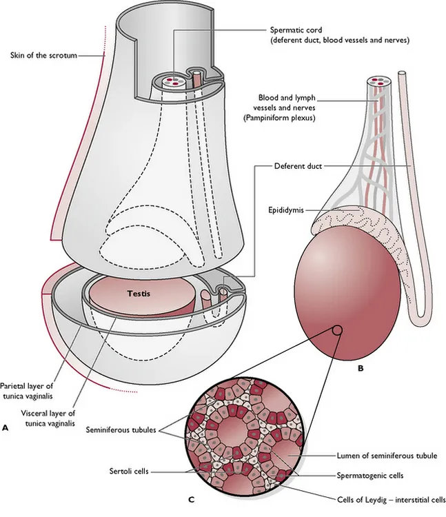
The testes are located outside the body cavity in the scrotum, a sac of hairless, often pigmented skin. This external positioning is crucial because spermatogenesis occurs most efficiently at temperatures slightly lower than the core body temperature. The scrotum is equipped with the dartos muscle, which contracts in cold weather to raise the testes closer to the body for warmth and relaxes in warm weather to cool them down.
Internally, each testis is wrapped in a double layer of peritoneum called the tunica vaginalis. The testicular tissue consists of seminiferous tubules, where spermatogenesis occurs. These tubules are lined with spermatogenic cells (which divide to produce sperm) and Sertoli cells (which secrete nutrients and estrogen to support sperm survival). Between the tubules lie the cells of Leydig, which secrete testosterone under the influence of interstitial cell-stimulating hormone (ICSH) from the pituitary gland.
Epididymis and Deferent Duct
Sperm produced in the seminiferous tubules are transported to the epididymis, a coiled tube located along the dorsolateral border of the testis. Here, sperm undergo maturation and are stored until ejaculation. The epididymis continues as the deferent duct (vas deferens), which carries sperm from the epididymis to the urethra during ejaculation.
The deferent duct passes through the inguinal canal as part of the spermatic cord, which also contains the testicular artery, vein, and nerve. The cremaster muscle within the spermatic cord contracts to raise the testis closer to the body in response to cold, working alongside the dartos muscle to maintain optimal temperature for spermatogenesis.
Penis: The Copulatory Organ
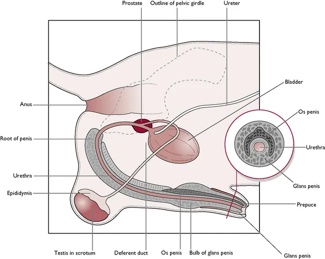
The penis serves dual functions: conveying sperm into the female reproductive tract during mating and expelling urine from the bladder. Its structure varies between dogs and cats.
In Dogs
The canine penis consists of:
- Corpus spongiosum penis: Erectile tissue surrounding the urethra, expanding into the bulb of the penis and the glans penis.
- Corpus cavernosum penis: Two crura (singular: crus) that attach the penis to the ischial arch of the pelvis.
- Os penis: A bone within the glans penis that aids in penetration during mating.
The penis is sheathed in the prepuce, a fold of skin that protects the glans. During mating, the prepuce retracts to expose the glans. The retractor penis muscle pulls the penis back into the prepuce after mating.
In Cats
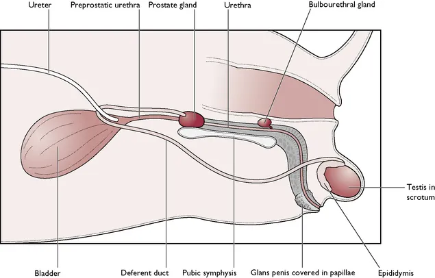
The feline penis is shorter and points backward, with the external opening located ventral to the anus. The glans penis is covered with tiny barbs, which stimulate ovulation in the female during mating—a process known as induced ovulation. The os penis in cats lies ventral to the urethra.
Accessory Glands
Accessory glands in the male reproductive system secrete seminal fluids, which:
- Increase the volume of the ejaculate to aid sperm transport.
- Provide a suitable environment for sperm survival.
- Neutralize the acidity of urine in the urethra.
The prostate gland is present in both dogs and cats and surrounds the urethra. In dogs, it is located near the neck of the bladder, while in cats, there is a short preprostatic urethra cranial to the gland. Bulbourethral glands are found only in tomcats and lie on either side of the urethra, cranial to the ischial arch.
Female Reproductive System
The female reproductive system in dogs (bitches) and cats (queens) is designed to produce ova, support fertilization, and nurture developing embryos. It consists of the following components:
Ovary: The Female Gonad
The ovary is the primary female reproductive organ, with two key functions:
- Production of ova (eggs) for fertilization by sperm.
- Endocrine function: Secretion of estrogen and progesterone, hormones that regulate the oestrous cycle and prepare the reproductive tract for pregnancy.
The ovaries are located in the dorsal abdominal cavity, caudal to the kidneys, and are held in place by the ovarian (suspensory) ligament. Each ovary is suspended from the dorsal body wall by the mesovarium, a fold of the visceral peritoneum that also encloses the infundibulum of the uterine tube. The ovarian bursa, a pocket-like structure within the mesovarium, covers the ovary and contains a small opening for ova to exit.
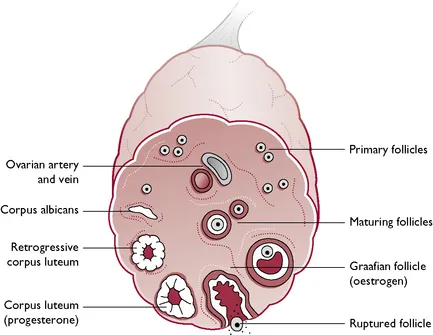 Longitudinal section of an ovary, showing follicles and germ cells
Uterine Tube (Oviduct or Fallopian Tube)
The uterine tube collects ova as they are released from the Graafian follicles and transports them to the uterine horns. Its open end, the infundibulum, is fringed with fimbriae, finger-like processes that capture ova from the ovary. The tube is lined with ciliated columnar epithelium, which propels the ova toward the uterus.
Uterus: The Site of Embryonic Development
The uterus is a Y-shaped structure consisting of two uterine horns and a central body. Its functions include:
- Providing a receptacle for embryonic development.
- Supporting the survival of embryos.
- Facilitating nutrient exchange between the dam and the developing embryos via the placenta.
The uterine wall has three layers:
- Endometrium: A glandular lining that thickens during pregnancy to nourish the embryo.
- Myometrium: Layers of smooth muscle that contract during parturition (birth).
- Mesometrium (Broad Ligament): A fold of the visceral peritoneum that suspends the uterus from the dorsal body wall.
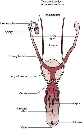
Cervix, Vagina, and Vestibule
- Cervix: A muscular sphincter connecting the uterine body to the vagina. It remains tightly closed except during mating or parturition, when it relaxes to allow the passage of sperm or fetuses.
- Vagina and Vestibule: These form a channel leading to the external opening of the reproductive tract, the vulva. The vagina extends from the cervix to the external urethral orifice, while the vestibule leads from the urethral orifice to the vulva. Both are lined with stratified squamous epithelium, which undergoes hormonal changes during the oestrous cycle.
Vulva and Mammary Glands
- Vulva: The external opening of the urogenital tract, consisting of the labia (vertical lips) and the clitoris (a structure of erectile tissue).
- Mammary Glands: Modified cutaneous glands essential for lactation, the production of milk to nourish newborns. Dogs have five pairs of mammary glands, while cats have four pairs. Milk production is influenced by hormones like progesterone, prolactin, and oxytocin.
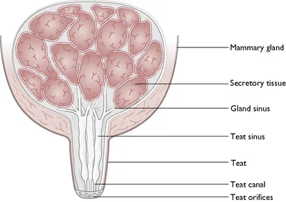 Section through a mammary gland, showing glandular tissue and teat canals
Lactation and Milk Composition
Lactation begins during pregnancy and is influenced by hormonal changes. The first milk produced, colostrum, is rich in maternal antibodies that provide passive immunity to newborns. Colostrum must be consumed within the first 24 hours of life, as the newborn’s intestine can only absorb antibodies during this window. After a few days, colostrum production stops, and the milk’s composition stabilizes.
The average composition of milk in dogs and cats includes:
- Water: 70–90%
- Fat: 0–30%
- Protein: 1–15%
- Carbohydrates: 3–7%
- Minerals: 1.5–1% (e.g., calcium, phosphorus, magnesium)
- Vitamins: A, B2, B5, E, K (low in vitamins C and D)
Notably, cat milk contains the amino acid taurine, which is essential for feline health.
The Oestrous Cycle
The oestrous cycle is a rhythmic series of hormonal and physiological changes in sexually mature, non-pregnant female mammals. It prepares the female for mating and pregnancy by:
- Producing ova ready for fertilization.
- Preparing the reproductive tract to receive fertilized ova.
- Initiating behavioral changes to signal receptivity to males.
The cycle includes a period of oestrus, during which the female is sexually receptive. In dogs, the oestrous cycle typically lasts about 6–7 months, while in cats, it is seasonal and influenced by environmental factors like daylight and temperature.
Key Genetic Concepts
Reproduction is deeply tied to genetics, the science of inheritance. Genetic information is carried on genes within the nucleus of every cell. During reproduction:
- Meiosis: Germ cells (sperm and ova) are produced with a haploid (half) set of chromosomes.
- Fertilization: A sperm and an ovum fuse to form a zygote, which undergoes cell division to develop into an embryo and then a fetus.
- Mendel’s Laws of Inheritance: These laws predict the outcomes of selective breeding by describing how traits are passed from parents to offspring.
Conclusion
The reproductive systems of dogs and cats are complex and highly specialized, ensuring the survival and perpetuation of their species. Understanding the anatomy, physiology, and hormonal regulation of these systems is essential for veterinarians, breeders, and pet owners alike. Whether it’s the production of sperm and ova, the intricacies of the oestrous cycle, or the nurturing of offspring through lactation, each component plays a vital role in the reproductive process.
For those interested in learning more, consulting veterinary resources or seeking guidance from a licensed veterinarian is always recommended.