The male reproductive system in both dogs and tomcats is a complex and fascinating biological system designed for the production and delivery of sperm, as well as the secretion of hormones crucial for male development and behavior. While sharing fundamental functions, there are notable anatomical distinctions between these two species. This article delves into the anatomy, function, and specific characteristics of the male reproductive tracts in dogs and tomcats, offering insights for pet owners and veterinary professionals alike.
Core Functions of the Male Reproductive System
The primary roles of the male reproductive system can be summarized as follows:
- Spermatogenesis: The production of spermatozoa, or sperm, which are essential for fertilizing the female ova. This process occurs optimally at temperatures below 40°C.
- Fluid Secretion: The production of seminal fluids that not only aid in sperm survival but also facilitate their transport into the female reproductive tract during mating.
- Hormone Production: The secretion of testosterone, a vital hormone responsible for the development of secondary sexual characteristics and influencing male behavior patterns.
Anatomical Structures and Their Functions
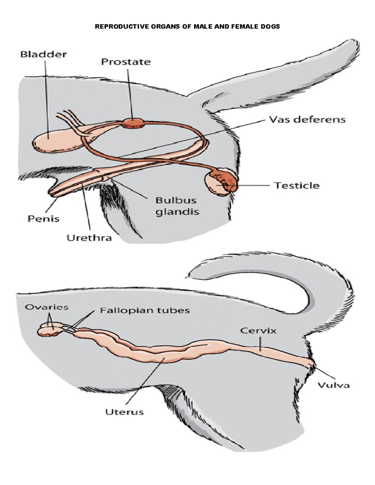
Testes and Scrotum
A pair of testes are responsible for sperm production. In adult dogs and tomcats, the testes are located outside the main body cavity within the scrotum, a pouch of pigmented, sparsely furred skin. The position of the scrotum differs: in dogs, it lies between the upper thighs, while in tomcats, it is situated ventral to the anus, near the ischial arch.
The scrotum’s structure plays a role in temperature regulation. The Dartos muscle within the scrotal skin contracts in cold weather, pulling the testes closer to the body for warmth, and relaxes in warm weather, allowing the testes to descend and cool.
Within the testes, coiled seminiferous tubules are lined with spermatogenic cells (producing sperm) and Sertoli cells (providing nutrients and secreting estrogen). Interstitial cells (Leydig cells), located between the tubules, are responsible for secreting testosterone.
Epididymis and Deferent Duct
The seminiferous tubules converge to form the epididymis, a long, coiled tube located along the dorso-lateral border of the testis. Here, sperm undergo final maturation and are stored. The epididymis continues as the deferent duct (vas deferens or ductus deferens), which ascends from the scrotum into the peritoneal cavity via the inguinal ring.
Urethra
The urethra serves as a shared passage for both the urinary and reproductive systems, extending from the bladder’s neck to the external opening at the tip of the penis. In dogs, the urethra is divided into a pelvic part and a longer penile part. In tomcats, there is a shorter preprostatic urethra, and the penile urethra is significantly shorter, with its opening directed caudally. This latter feature is associated with the tomcat’s territorial marking behavior.
Accessory Glands
Both dogs and tomcats possess a prostate gland near the bladder neck. Additionally, tomcats have bulbo-urethral glands located near the tip of the penis. These glands contribute secretions that increase ejaculate volume, neutralize acidity in the urethra, and create a favorable environment for sperm.
Penis
The penis contains erectile tissue (corpus cavernosum penis) that engorges with blood during sexual excitement, enabling intromission. Significant differences exist between the canine and feline penis.
- Dog: The dog’s penis has a glans penis that forms about a quarter of its length and contains the os penis, a small bone that aids rigidity during the initial stages of mating. The urethra runs through a bony tunnel within the os penis, which can be a site for blockages.
- Tomcat: The tomcat’s penis has a bony os penis within the erectile tissue, where the urethra is narrow and prone to blockage by crystals. The glans penis is covered in small, backward-pointing barbs. These barbs induce pain upon withdrawal, initiating a reflex ovulation in the female (queen) approximately 36 hours later, characteristic of induced ovulators.
Prepuce
When relaxed, the penis is enclosed within the prepuce, a protective sheath lined with mucous membrane. Infections of the prepuce, known as balanoprosthitis, can result in an unpleasant discharge.
Understanding these anatomical and functional differences is crucial for appreciating the reproductive biology of these common companion animals.
References:
- ASPINALL, V. and CAPELLO, M, 2009. Introduction to Veterinary Anatomy and Physiology. 2nd ed. Butterworth Heinemann. Oxford.
- DYCE, K. M., SACK, W. 0. and WENSING. C. J. G., 2002. Textbook of Veterinary Anatomy. 3rd ed. Saunders. Philadelphia.
- EVANS. H. E., 1993. Miller’s Anatomy of the Dog. 3rd ed. Saunders. Philadelphia.
- Veterinary Nursing Journal • VOL 26 • March 2011 •